BASIC INFORMATION
Date & Time: June 8, 2026, 20:22:01 Indian Standard Time
Lecture Handout Prepared from the Teaching Session by: Dr. R. K. Mishra
SUMMARY
This lecture provides a comprehensive overview of small bowel obstruction (SBO), from fundamental gastrointestinal physiology to advanced management strategies. It begins by explaining the highly regulated processes of motility, secretion, and neurohormonal control, which are catastrophically disrupted in SBO. The pathophysiology of obstruction is detailed, emphasizing the progression from simple luminal blockage to the life-threatening vascular compromise seen in strangulated and closed-loop variants. A significant portion of the lecture is dedicated to the etiology of SBO, focusing on postoperative adhesions, which account for the majority of cases. The biological cascade of adhesion formation—from peritoneal injury and inflammatory signaling to fibrinolytic failure—is thoroughly explained. The lecture also covers high-acuity emergent variants, such as internal hernias, particularly those arising as a late complication of Roux-en-Y gastric bypass surgery. Diagnostic protocols are outlined, contrasting traditional plain radiography with the gold standard, contrast-enhanced CT, and highlighting critical radiological signs and the importance of serum lactate in assessing bowel viability. Finally, the lecture delineates management pathways, including the principles of non-operative management with Gastrografin, absolute indications for surgery, a comparison of laparoscopic versus open adhesiolysis, and evidence-based strategies for adhesion prevention.
KEY KNOWLEDGE POINTS
-
Baseline gastrointestinal physiology involves integrated endocrine, paracrine, and neural (extrinsic and intrinsic enteric) control systems.
-
The four cardinal symptoms of obstruction are colicky abdominal pain, nausea and vomiting, abdominal dilation, and obstipation.
-
SBO is classified by etiology (intraluminal, intramural, extrinsic), degree (partial/complete), and progression (simple/strangulated).
-
Rising intraluminal pressure sequentially collapses venous outflow then arterial inflow, leading to ischemia, necrosis, and perforation.
-
Postoperative adhesions are the dominant cause of SBO (~56%), resulting from peritoneal injury, cytokine-driven inflammation, and fibrinolytic failure (decreased TPA, increased PAI).
-
Closed-loop obstruction, internal hernias, and volvulus are surgical emergencies due to rapid vascular compromise, with strangulation possible in as little as six hours.
-
Roux-en-Y gastric bypass is a leading cause of acquired internal hernias due to iatrogenic mesenteric defects that widen with postoperative weight loss.
-
Contrast-enhanced CT is the gold standard for diagnosis; oral contrast is contraindicated due to aspiration risk and diagnostic obscuration.
-
Key CT signs include the transition point, whirl sign (volvulus), beak sign, and small bowel feces sign; signs of ischemia include reduced wall enhancement and pneumatosis intestinalis.
-
Serum lactate is a critical biochemical marker for bowel ischemia.
-
Non-operative management for adhesive SBO includes bowel rest, NG decompression, and the therapeutic use of water-soluble hyperosmolar contrast (Gastrografin).
-
Laparoscopic adhesiolysis, when feasible, improves short-term outcomes over open surgery but carries a notable risk of enterotomy and conversion.
-
Adhesion prevention focuses on meticulous tissue handling, minimal thermal injury, and barrier agents like Seprafilm, which must not be placed near anastomoses.
INTRODUCTION
The human gastrointestinal tract is a structurally elaborate, twenty-foot-long biological system designed for complex macromolecule assimilation, requiring absolute physiological synchrony. Small bowel obstruction (SBO) represents a common and potentially fatal surgical emergency, characterized by a catastrophic disruption of this transit system. While a mechanical blockage initiates the syndrome, the lethal trajectory is driven by escalating intraluminal pressure, compromised microcirculation, systemic derangements, and, in severe cases, strangulation. The closed-loop variant is a distinct and highly lethal entity where a bowel segment is occluded at two points, leading to rapid gangrene and perforation with a mortality rate up to 35%. Adhesions, primarily postoperative, are the dominant etiology. Understanding the pathology, from baseline physiology to the biology of peritoneal healing and the nuances of high-risk variants, is essential for prompt diagnosis, effective prevention, and sound surgical decision-making.
LEARNING OBJECTIVES
-
To comprehend the physiological mechanisms of gastrointestinal motility, secretion, and neurohormonal regulation.
-
To classify small bowel obstructions and describe the sequential pathophysiological events leading to ischemia, necrosis, and perforation.
-
To explain adhesion formation, focusing on mesothelial injury, cytokine signaling, and fibrinolytic failure.
-
To recognize the clinical urgency of high-risk variants, including closed-loop obstruction, internal hernias (congenital and acquired), and volvulus.
-
To identify and interpret key clinical, radiological, and biochemical signs of SBO and bowel strangulation.
-
To understand the principles of non-operative management, including the role of water-soluble contrast, and delineate absolute indications for surgical intervention.
CORE CONTENT
1. Normal Gastrointestinal Physiology
The primary objective of the GI tract is nutrient assimilation via four overlapping processes:
-
Motility: Mechanical grinding, churning, and forward propulsion of luminal contents.
-
Secretion: Continuous influx of water, electrolytes, and digestive enzymes.
-
Digestion: Chemical degradation of unabsorbable macromolecules.
-
Absorption: Transport of microscopic nutrients across the intestinal epithelium.
1.1. Regulatory Control Systems
-
Endocrine System: Mediated by "open-type" enteroendocrine cells that sample luminal contents (e.g., pH, fats) and release systemic hormones like cholecystokinin (CCK).
-
Paracrine System: Provides localized control via interstitial diffusion of messengers like histamine and somatostatin.
-
Neural System:
-
Extrinsic Control: The autonomic nervous system, with parasympathetic (vagal) stimulation enhancing digestion and sympathetic activity inhibiting it.
-
Intrinsic Control (Enteric Nervous System): An independent, dense neuronal network within the bowel wall, comparable in neuron count to the spinal cord, that autonomously coordinates peristalsis.
-
1.2. Intestinal Fluid Dynamics
A fluid environment is maintained by the osmotic pull of water following the active secretion of electrolytes (sodium, chloride) into the lumen.
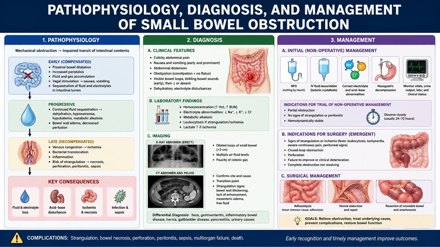
2. Pathophysiology of Small Bowel Obstruction (SBO)
SBO is the failure of aboral propulsion of intestinal contents.
-
Functional Obstruction (Ileus): Paralysis of the intestinal neuromuscular machinery without a physical blockage, caused by inflammation, infection, or ischemia.
-
Mechanical Obstruction: Accounts for over 80% of cases and involves a physical barricade. The four cardinal symptoms are colicky abdominal pain, nausea and vomiting, abdominal dilation, and obstipation.
-
Clinical Pathophysiology:
-
Colicky Pain: The enteric nervous system mounts intense hyperperistaltic contractions against the fixed blockage.
-
Vomiting and Distension: Upstream accumulation of fluid, gas, and secretions leads to vomiting and bowel dilation.
-
Vascular Compromise: Rising intraluminal pressure first collapses low-pressure venous outflow, causing congestion and edema. This further elevates pressure until arterial inflow is compromised, leading to ischemia, necrosis, and perforation.
-
Systemic Effects: Vomiting and fluid sequestration cause dehydration, hypokalemia, hypochloremia, and reduced intravascular volume.
-
3. Classification and Etiology of Mechanical Obstruction
3.1. Classification by Etiology
-
Intraluminal: Obstruction within the lumen (e.g., gallstone ileus, bezoars, foreign bodies).
-
Intramural: Obstruction originating in the bowel wall (e.g., Crohn's strictures, neoplasms).
-
Extrinsic: Compression from outside the bowel (e.g., adhesions, hernias, tumors). This is the most common category.
3.2. Classification by Progression
-
Simple Obstruction: Luminal transit is blocked, but vascular supply is intact.
-
Strangulated Obstruction: Vascular supply is compromised, leading to ischemia.
3.3. Etiology and Epidemiology
-
Adhesions: ~56% (dominant cause).
-
Hernias: ~25%.
-
Tumors: ~10%.
-
Inflammatory diseases: ~9%.
4. Postoperative Adhesion Formation
-
Mesothelial Injury: The peritoneum is lined by a fragile single layer of mesothelial cells easily damaged by friction, handling, and air exposure.
-
Inflammatory Cascade: Injury recruits macrophages, which release IL-1 and TNF, inducing adhesionogenic IL-6. Local hypoxia drives VEGF expression, and TGF-β stimulates fibroblast proliferation.
-
Fibrinolytic Failure: Normal healing involves temporary fibrin deposition followed by lysis. In adhesion formation, mesothelial cell loss reduces local tissue plasminogen activator (TPA) production, while inflammation increases plasminogen activator inhibitor (PAI). This imbalance prevents fibrin dissolution.
-
Scar Maturation: The persistent fibrin scaffold is infiltrated by fibroblasts, which deposit collagen. VEGF-supported neovascularization stabilizes the structure into a permanent, fibrous adhesion band.
-
Evolutionary Context: Adhesions are adaptive in traumatic injury (plugging holes, containing contamination) but are maladaptive after sterile surgery.
5. Emergent Variants: Closed-Loop Obstruction, Internal Hernias, and Volvulus
-
Closed-Loop Obstruction: A bowel segment is occluded at two points, creating a sealed chamber. This prevents decompression, leading to a rapid rise in intraluminal pressure and accelerated vascular collapse. Gangrene can develop in as little as six hours.
-
Internal Hernias: Protrusion of bowel through a congenital or acquired defect within the peritoneal cavity. They are a rare cause of SBO (<1%) but have a high strangulation risk.
-
Congenital: Arise from embryological defects (e.g., paraduodenal hernias through the fossa of Lanzert, transmesenteric defects).
-
Acquired: Most commonly iatrogenic, with a rising incidence due to Roux-en-Y gastric bypass (RYGB) surgery. Surgically created mesenteric defects, though sutured, can reopen and widen as massive postoperative weight loss causes resorption of mesenteric fat.
-
-
Volvulus: Twisting of the bowel on its mesentery, which acts as a tourniquet, acutely compromising the vascular pedicle.
-
Clinical Presentation of Internal Hernias: Can be notoriously variable, from chronic, intermittent, reducible hernias (often misdiagnosed as IBS) to acute, incarcerated presentations requiring immediate surgery.
6. Diagnosis and Initial Management
6.1. Diagnostic Imaging
-
Plain Radiography: Traditional first step. The classic triad includes:
-
Dilated small bowel loops (>3 cm).
-
Multiple air-fluid levels.
-
Paucity of gas in the colon.
-
The "string of pearls sign" (gas trapped between valvulae conniventes in a fluid-filled loop) is a specific sign.
-
Limitations: Low sensitivity, especially for closed-loop obstructions, which may be fluid-filled and radiographically occult.
-
-
Contrast-Enhanced Computed Tomography (CT): The gold standard (accuracy >95%).
-
Protocol: IV contrast is mandatory to assess bowel wall viability. Oral contrast is contraindicated due to aspiration risk and its potential to obscure wall enhancement.
-
Key Findings:
-
Transition Point: The site where dilated proximal bowel collapses to a decompressed caliber.
-
Small Bowel Feces Sign (SBFS): Particulate material in the dilated small bowel, located just proximal to the transition point.
-
Signs of Closed-Loop/Volvulus: U- or C-shaped loop, beak sign (tapering at the point of torsion), and whirl sign (swirling of mesenteric vessels).
-
-
Signs of Strangulation/Ischemia:
-
Reduced or absent bowel wall enhancement.
-
Bowel wall thickening (edema).
-
Mesenteric fat stranding and free fluid (ascites).
-
Pneumatosis Intestinalis: Gas in the bowel wall (a grave sign of necrosis).
-
Portal Venous Gas: A pre-terminal sign with extremely high mortality.
-
-
6.2. Laboratory Investigations
-
Complete Blood Count (CBC): Leukocytosis may indicate stress or inflammation.
-
Basic Metabolic Panel: Assesses for dehydration and electrolyte abnormalities (hypokalemia, hypochloremia).
-
Serum Lactate: A vital marker for bowel ischemia. A rising lactate level is a highly sensitive indicator mandating urgent surgery.
6.3. Non-Operative Management (NOM) for Adhesive SBO
Indicated for stable patients with presumed adhesive SBO without signs of strangulation.
-
Principles: Bowel rest (NPO), nasogastric (NG) tube decompression, and IV fluid resuscitation.
-
Time Limits: Conservative management should not exceed 72 hours without resolution.
-
The Gastrografin Challenge: Administration of a hyperosmolar water-soluble contrast agent (e.g., Gastrografin).
-
Mechanism: Draws water from the edematous bowel wall via osmosis, reducing edema. The influx of fluid also stimulates a strong peristaltic wave.
-
Role: Can be both therapeutic (accelerating resolution) and diagnostic (contrast reaching the colon on a 24-hour X-ray predicts success).
-
7. Operative Management
7.1. Indications for Immediate Surgery
-
Absolute Indications: Peritonitis, hemodynamic instability, or CT signs of strangulation (pneumatosis, portal venous gas, closed-loop obstruction, definite necrosis).
-
Predictors of NOM Failure: Presence of multiple factors such as free intraperitoneal fluid, severe mesenteric edema, and intractable vomiting supports earlier operative intervention.
-
Virgin Abdomen: An SBO in a patient with no prior abdominal surgery warrants expedited exploration, as a non-adhesive cause (e.g., tumor, internal hernia) is likely.
7.2. Operative Strategy: Adhesiolysis
-
Laparoscopic Approach: Preferred when feasible. Associated with shorter operative time, shorter hospital stay, and lower mortality compared to open surgery. However, it carries a ~7% risk of iatrogenic enterotomy and a conversion rate to open surgery of ~29%.
-
Open Laparotomy: Indicated for patients with dense adhesions, massively dilated bowel, or when laparoscopic access is unsafe.
8. Prevention of Adhesions
-
Meticulous Surgical Technique: Gentle tissue handling, minimizing thermal injury from electrocautery, and copious irrigation with warm saline to remove blood and debris. Laparoscopy is preferred to limit mesothelial trauma.
-
Pharmacologic Strategies: Largely unsuccessful due to systemic side effects. Antifibrotics (e.g., halofuginone) and anti-inflammatories (NSAIDs) impair necessary wound and anastomotic healing. Fibrinolytics (e.g., tPA) cause significant hemorrhage.
-
Barrier Agents:
-
Seprafilm: A bioresorbable membrane of hyaluronic acid and carboxymethylcellulose that creates a temporary physical barrier between surfaces. It reduces adhesion severity but must not be placed near an anastomosis, as it may increase the risk of anastomotic leak and mortality.
-
-
Historical Procedures: Plication techniques (e.g., Noble's plication), which involved suturing the bowel into orderly folds, are now obsolete due to high complication rates.
SURGICAL PEARLS
-
The primary pitfall in managing SBO is failure to recognize a strangulated bowel. Differentiating simple from strangulated obstruction is the immediate clinical priority.
-
A rising serum lactate in a patient with SBO is a surgical emergency; the biochemical signal often precedes definitive radiological signs of necrosis.
-
In a post-bariatric bypass patient with abdominal pain, internal hernia must be a primary differential diagnosis until proven otherwise, even years after surgery and with a "normal" CT scan.
-
Never administer oral contrast for an initial diagnostic CT in suspected SBO; it risks aspiration and obscures bowel wall enhancement.
-
The small bowel feces sign is a reliable pointer on CT, indicating you are immediately proximal to the site of obstruction.
-
Gentle, deliberate tissue handling, minimal cautery, and copious irrigation are the cornerstones of adhesion prevention.
ANESTHETIC AND PHYSIOLOGICAL CONSIDERATIONS
-
The failure of aboral transit coupled with continued active ion and fluid secretion leads to massive intraluminal fluid sequestration.
-
Clinicians must anticipate severe dehydration, third-spacing of fluids, and significant electrolyte abnormalities (such as hypochloremia and hyponatremia) requiring aggressive preoperative resuscitation.
COMPLICATIONS AND THEIR MANAGEMENT
-
Intraoperative:
-
Iatrogenic enterotomy during adhesiolysis requires meticulous technique to avoid, especially with dilated, edematous bowel. If it occurs, it may necessitate conversion to open laparotomy, peritoneal washout, and segmental resection.
-
-
Early Postoperative:
-
Prolonged ileus, anastomotic leak (risk heightened by misapplied barrier agents), and progression of unrecognized ischemia leading to necrosis, perforation, and peritonitis.
-
-
Late Postoperative:
-
Adhesions are the leading cause of SBO and may lead to recurrent mechanical obstructions long after the index operation.
-
Internal hernias are a significant late complication of RYGB.
-
MEDICOLEGAL AND PATIENT SELECTION CONSIDERATIONS
-
Failure or delay in diagnosing a strangulated obstruction is a significant source of morbidity, mortality, and medicolegal liability.
-
The decision to pursue non-operative management must be based on a stable patient with no signs of strangulation, and the rationale must be rigorously documented. Adherence to time limits (e.g., 72 hours) is critical.
-
Informed consent for RYGB must include the risk of late complications like internal hernia. Misdiagnosing intermittent symptoms in these patients as functional can lead to significant harm.
-
Informed consent for adhesiolysis must include the risks of enterotomy, conversion to open surgery, and recurrence.
SUMMARY AND TAKE-HOME MESSAGES
-
The gastrointestinal tract is highly regulated by simultaneous endocrine, paracrine, and enteric nervous system pathways to ensure nutrient assimilation.
-
SBO evolves via a pressure-driven vascular failure cascade: venous collapse precedes arterial compromise, culminating in ischemia and necrosis.
-
Over 80% of bowel obstructions are mechanical, with postoperative adhesions being the most frequent cause, driven by fibrinolytic failure.
-
Computed tomography with IV contrast is the gold standard for diagnosis; oral contrast is contraindicated. Serum lactate is a key marker for ischemia.
-
The transition from simple to strangulated obstruction, or the presence of a closed-loop variant, marks a critical, life-threatening progression that requires emergent surgical intervention.
-
Laparoscopic adhesiolysis offers superior short-term outcomes but has significant risks; prevention through meticulous surgical technique is paramount.
MULTIPLE CHOICE QUESTIONS (MCQs)
-
Which regulatory system of the gastrointestinal tract possesses intrinsic autonomy with a neural density comparable to the spinal cord?
A) Endocrine System
B) Paracrine System
C) Extrinsic Autonomic Nervous System
D) Intrinsic Enteric Nervous System
Answer: D
-
The most common etiology of mechanical small bowel obstruction is:
A) Intraluminal bezoars
B) Intramural tumors
C) Extrinsic adhesions
D) Functional ileus
Answer: C
-
What is the critical distinction between a simple and a strangulated mechanical obstruction?
A) A simple obstruction is partial, while a strangulated obstruction is complete.
B) A simple obstruction has an intact blood supply, whereas a strangulated obstruction features compromised vascular flow.
C) A simple obstruction presents with obstipation, while a strangulated one does not.
D) A simple obstruction is caused by adhesions, while a strangulated one is caused by a hernia.
Answer: B
-
The pathophysiological mechanism of adhesion formation is primarily driven by:
A) Excessive production of tissue plasminogen activator (TPA).
B) Failure of fibrinolysis due to decreased TPA and increased plasminogen activator inhibitor (PAI).
C) An anti-inflammatory response mediated by IL-10.
D) Rapid re-epithelialization of the entire peritoneal surface.
Answer: B
-
What is the defining characteristic of a closed-loop bowel obstruction?
A) Obstruction caused by a malignant tumor.
B) Obstruction of the bowel at two distinct points.
C) Obstruction accompanied by profuse watery diarrhea.
D) Obstruction that resolves spontaneously within 6 hours.
Answer: B
-
The rising incidence of acquired internal hernias is most closely associated with which surgical procedure?
A) Appendectomy
B) Open cholecystectomy
C) Roux-en-Y gastric bypass
D) Colectomy for cancer
Answer: C
-
What is the primary reason for avoiding oral contrast during the initial CT scan for a suspected SBO?
A) It is too expensive for an emergency setting.
B) It can cause an allergic reaction.
C) It obscures the view of IV-enhanced bowel wall and poses an aspiration risk.
D) It is less effective than IV contrast at showing dilation.
Answer: C
-
Which CT sign is definitive for a volvulus?
A) The beak sign
B) The small bowel feces sign
C) The whirl sign
D) The string of pearls sign
Answer: C
-
A rising serum lactate in a patient with SBO is most indicative of:
A) Dehydration
B) Bowel ischemia
C) Hypokalemia
D) Normal physiological stress
Answer: B
-
The therapeutic effect of Gastrografin in adhesive SBO is primarily due to its:
A) Pro-kinetic properties.
B) Lubricating effect.
C) Antibacterial action.
D) Hyperosmolar nature, which draws water from the edematous bowel wall.
Answer: D
-
What is the most severe and prognostically grave radiological sign in a patient with SBO?
A) Bowel wall thickening
B) Mesenteric fat stranding
C) The small bowel feces sign
D) Portal venous gas
Answer: D
-
A patient with a "virgin abdomen" (no prior surgery) presents with SBO. What is the most appropriate management step?
A) A prolonged trial of non-operative management for 96 hours.
B) Immediate administration of Gastrografin.
C) Expedient surgical exploration due to the high likelihood of a non-adhesive etiology.
D) Discharge with a diagnosis of gastroenteritis.
Answer: C
-
Which of the following is an absolute indication for immediate surgery in a patient with SBO?
A) A white blood cell count of 12,000/mm³.
B) A CT scan showing a clear transition point without ischemic signs.
C) Peritonitis on physical examination.
D) Mild abdominal distension.
Answer: C
-
Compared to open adhesiolysis, the laparoscopic approach is associated with:
A) A longer hospital stay.
B) A higher 30-day mortality rate.
C) A shorter operative time and hospital stay.
D) A zero percent risk of enterotomy.
Answer: C
-
What is the key limitation of using systemic anti-inflammatory (NSAIDs) or antifibrotic agents to prevent adhesions in humans?
A) They are prohibitively expensive.
B) They impair necessary wound and anastomotic healing.
C) They are only effective when given preoperatively.
D) They cause severe renal toxicity.
Answer: B
-
A left paraduodenal hernia occurs through a congenital defect known as:
A) The foramen of Winslow
B) The pouch of Douglas
C) The fossa of Lanzert
D) Morison's pouch
Answer: C
-
The "small bowel feces sign" on a CT scan is clinically useful because it:
A) Confirms the patient has a fecal impaction.
B) Indicates the obstruction is located in the colon.
C) Is almost always located immediately proximal to the transition point.
D) Is a definitive sign of bowel perforation.
Answer: C
-
A key risk of applying a barrier agent like Seprafilm is:
A) Increased risk of anastomotic leak if placed across a suture line.
B) Severe allergic reaction in most patients.
C) Paradoxical increase in dense adhesions.
D) Intra-abdominal abscess formation in all cases.
Answer: A
-
Which of the following is NOT one of the four cardinal symptoms of mechanical bowel obstruction?
A) Colicky abdominal pain
B) Abdominal dilation
C) Profuse watery diarrhea
D) Nausea and vomiting
Answer: C
-
The initial vascular event as intraluminal pressure rises in an obstructed bowel loop is:
A) Arterial inflow compromise.
B) Capillary rupture.
C) Venous outflow collapse.
D) Lymphatic thrombosis.
Answer: C
MOTIVATIONAL MESSAGE FROM DR. R. K. MISHRA
The path to surgical mastery is paved not with haste, but with deliberate practice. Each moment of study, each repetition of technique, sharpens the judgment that will one day protect a life.
Wishing you the fortitude to learn relentlessly and the wisdom to act decisively. Your dedication today shapes the future of medicine.
| Older Post | Home | Newer Post |


