Ureteral stenting
Definition
A ureteral stent is really a thin, flexible tube threaded to the ureter to assist urine drain in the kidney towards the bladder into another collection system.
Description
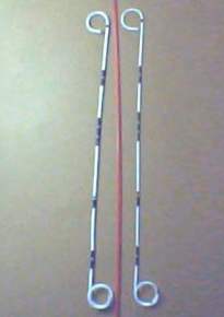
The dimensions, shape, and material from the ureteral stent for use rely on the patient's anatomy and also the reason why the stent is needed. Most stents are 12-30 cm long, and also have a diameter of just 1.5-6 mm. Either ends from the stent might be coiled known as a pigtail stent to avoid it motionless unnatural; an open-ended stent is much better suited to patients who need temporary drainage. Sometimes, one end from the stent includes a thread mounted on it that extends with the bladder and urethra towards outside the body; this helps with stent removal. The stent material should be flexible, durable, non-reactive, and radiopaque.
The individual is generally placed directly under general anesthesia for stent insertion; this makes certain problems how the patient will stay relaxed and won't move throughout the procedure. A cystoscope is really a thin, telescope-like device, is inserted to the urethra towards the bladder, and also the opening towards the ureter to become stented is identified. Sometimes, helpful information wire is inserted to the ureter underneath the aid of the fluoroscope. The guide wire supplies a path for that keeping the stent that is advanced within the wire. When the stent is within place, the guide wire and cystoscope are taken out. Patients who fail this process of ureteral stenting might have the stent positioned percutaneously, to the kidney, and subsequently to the ureter. A stent which has an attached thread might be brought out with a physician within an office setting. Cystoscopy could also be used to get rid of a stent.
Purpose
Urine is usually carried in the kidneys towards the bladder using a set of long, narrow tubes known as ureters by which each kidney is attached to one ureter. A ureter can become blocked due to numerous conditions as well as kidney stones, tumors, thrombus, postsurgical swelling, or infection. A ureteral stent is positioned within the ureter to revive the flow of urine towards the bladder. Ureteral stents can be utilized in patients with active kidney infection or with diseased bladders. Alternatively, ureteral stents can be utilized during or after urinary tract surgical treatments use a mold around which healing may appear, to change the urinary flow from regions of leakage, to control kidney stones or stop stone migration just before treatment, in order to result in the ureters easier identifiable in the course of difficult surgical treatments. The stent may stay to put on a short-term or long-term basis.
Demographics
Chronic blockage of the ureter affects around five individuals of all the 1,000; acute congestion affects one inch every 1,000. Bilateral blockage is rarer; chronic obstruction affects one person per 1,000 people, and acute blockage impacts five per 10,000.
Who performs the process and where could it be conducted?
Ureteral stenting is usually performed inside a hospital by an interventional radiologist is a physician who focuses on treating medical disorders using particular imaging techniques or perhaps a urologist is a physician who focuses on diagnosing and treating diseases from the urinary tract and genital organs.
Diagnosis/Preparation
A variety of technologies assisted in the proper diagnosis of ureteral blockage. Included in this are:
- ultrasonography is definitely an imaging technique that utilizes high-frequency sounds waves to visualize structures within the body
- cystoscopy is really a procedure when a thin, tubular device can be used to visualize the inside of the bladder
- pyelography is really a x-rays taken from the urinary tract following a contrast dye continues to be injected right into a vein or to the kidney, ureter, or bladder
- computed tomography is definitely an imaging technique that utilizes x-rays to create two-dimensional cross-sections on the viewing screen
Just before ureteral stenting, the process ought to be completely explained with a healthcare professional. No food or drink is allowed after midnight the night time before surgery. The individual wears a hospital gown throughout the procedure. When the stent insertion is conducted with a cystoscope, the individual will assume a situation that's typically utilized in a gynecological exam when a patient is lying about the back, using the legs flexed and based on stirrups.
Questions a patient should ask a doctor:
- What kind of stent is going to be used, so when could it be removed?
- Why is ureteral stenting suggested?
- Are there any options to ureteral stenting?
- What technique is going to be accustomed to put the stent?
- What diagnostic test is going to be performed before the stenting procedure?
Risks
Complications related to ureteral stenting include:
- bleeding is generally minor and easily treated, but from time to time needing transfusion
- catheter migration or dislodgement this might need readjustment
- coiling from the stent inside the ureter this might cause lower abdominal pain or flank pain on urination, urinary frequency, or blood within the urine
- introduction or deteriorating of infection
- penetration of adjacent organs
Normal results
Ordinarily, a ureteral stent re-establishes the circulation of urine in the kidney towards the bladder. Postoperative the flow of urine is going to be monitored to guarantee the stent is not dislodged or obstructed.
Morbidity and mortality rates
Serious complications exist in around 4% of patients going through ureteral stenting, with minor problems in another 10%.
Aftercare
Stents should be periodically replaced to avoid fractures inside the catheter wall or build-up of encrustation. Stent alternative is recommended around every 6 months; more regularly in patients who form stones.
Alternatives
If patient’s ureter is blocked and ureteral stenting isn't feasible, a nephrostomy might be performed. In this procedure, a tube is positioned with the skin about the patient's back, to the part of the kidney that accumulates urine. The tube might be attached to another drainage bag. In some cases, the tube is connected from the kidney towards the bladder.


