Assistant Professor/Lecturer
KIMS, Bangalore
Task Analysis of Laparoscopic Splenectomy
Indications
- Idiopathic Thrombocyotopenic Purport
- Autoimmune Haemolytic Anemia
- Microspherocytosis
- Benign tumours and cysts
- AIDS-related thrombocytopenia
Contra-Indications
- Massive Splenomegaly
- Portal Hypertension
- Vaccines - Pneumococcal , Haemophilus influenza, Neisseria meningitidis ideally two weeks prior to surgery or post operatively.
- Blood and platelet transfusion if needed and arrange blood.
Anaesthesia
- General anaesthesia with endotracheal intubation is required.
- Two large IV catheters.
- Foleys catheter and Nasogastric tube.
Patient Position
- Patient placed in right lateral position and left arm crossing chest and lying on right arm.
- Left hip and chest are elevated with pillows, leaving the flank area open and the left knee flexed, with a padding o blankets between the legs.
- The patients secured across the chest and hips to the table with wide adhesive tape,as the operating room table will be tilted.
- Monopolar cautery check done along with patient end plate and Hormonic scalpel checks done.
- Coaxial alignment of surgeon, target and monitor checked.
- Camera connected, white balancing and focusing done.
- Co2 cylinder checked for sufficient insuflation.
- Working status of all the lap instruments along with insulation check is properly made.
Operative Preparation
The skin is prepared from the lower chest to pubis.
Port placement
- A 10 mm camera port is inserted at the level of the umbilicus over the left mid clavicular line.
- A 2mm stab incision is placed and verses needle is inserted perpendicular to abdominal wall.
- Intrabdominal position if verses is confirmed by the suction, irrigation, hanging drop and plunger test.
- Pneumoperitoneum is created by setting the insufflator at 14 mm hg.
- Camera inserted and abdomen inspected noting the size of the spleen for working port placement.
- Two additional 5mm ports are inserted on either side of camera port at 7.5 cms according to base ball diamond concept.
- Additional epigastric port can be inserted for liver retraction in case of hepatomegaly.
Details of procedure
Dissecting free from ligaments

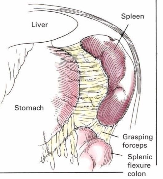
- After inspection of the abdomen the splenocolic ligament is visualised along with greater omentum.
- Splenic end of the ligament is identified and elevated with traction identifying a plane above the splenic flexure and entered using harmonic scalpel.
- Dissection continued medial to spleen to reach the gastrosplenic ligament containing short gastric vessels.
- By giving traction over the greater curvature of stomach lesser sac is entered using blunt dissection and short gastric vessels are divided 1 cm away from the gastric wall.
- The pancreas,splenic artery and vein running at the base of lesser sac are visualized.
- Short gastric are divided upto gastro oesophageal junction.
- Spleen is elevated medially and the splenorenal ligament is divided and continued till the top of spleen is free.

Dissection of splenic pedicle
- Dissection of the medial part of spleen continued to reach the splenic pedicle.
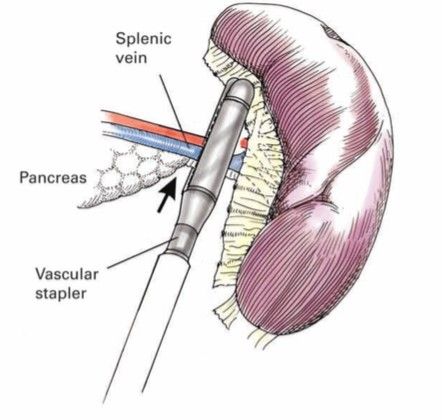
- The area chosen should be distal to the tail of the pancreas but proximal to the trifurcation of the splenic vessels.
- A 12 mm port is required for Endo GIA Vascular stapler for the splenic pedicle.
- Dissection is performed until vessels an be safely encompassed within the jaws of the Vascular stapler.
- Care is taken to include the pedicle having splenic artery and vein in the arms of the stapler and fired .
- Alternatively artery and vein can be dissected and fired with stapler separately.
- Reinforced plastic bag is introduced and the organ is carefully placed into the bag.
- Bag is closed and partially withdrawn through the abdominal wall until the open rim of the bag is under control outside the abdomen.
- Bag is cut free from the carrier using drawstring in the end of instrument handle.
- Spleen is morcellated and extracted
- Post extraction the right upper quadrant lavaged with suction irrigator and a careful inspection is made of all cut surfaces and vessels.
- Tail of the pancreas is examined for possible injury.
- A silastic catheter drain is placed.
- All ports are closed under vision.

Post operative care
- The NG tube is removed post operatively.
- Foleys catheter is discontinued when the patient is alert enough to void.
- Clear liquids can be started within a day and diet is advanced as tolerated.
beautifully explained and well presented lecture, video presentation and Task Analysis help me so much! Thank you!!
| Older Post | Home | Newer Post |
How to Perform and Implement Task Analysis of Laparoscopic and Robotic Procedures
Task analysis is a critical component of any complex surgical procedure, including laparoscopic and robotic surgeries. It involves breaking down the procedure into its constituent tasks, identifying the steps, skills, and cognitive processes required. Task analysis not only enhances the understanding of these intricate surgeries but also serves as a foundation for training, skill assessment, and continuous improvement in healthcare. In this essay, we will delve into how to conduct and implement task analysis for laparoscopic and robotic procedures.
Understanding the Significance of Task Analysis
Before we explore the procedure for task analysis, it's essential to recognize why it is of paramount importance in the realm of surgery, particularly for laparoscopic and robotic procedures.
1. Enhanced Learning and Training: Task analysis helps in developing structured training programs. It breaks down complex procedures into manageable components, making it easier for trainees to learn and practice each step methodically.
2. Skill Assessment: By understanding the tasks and sub-tasks involved, it becomes possible to assess the competence of surgeons and surgical teams. This is crucial for ensuring patient safety and quality care.
3. Workflow Optimization: Task analysis can reveal inefficiencies in surgical workflows. Identifying these bottlenecks allows for process improvements, potentially reducing surgical times and enhancing outcomes.
4. Error Reduction: Recognizing potential points of error is vital for preventing surgical complications. Task analysis can highlight critical steps where errors are more likely to occur, leading to proactive measures to mitigate risks.
Procedure for Task Analysis of Laparoscopic and Robotic Procedures:
Task analysis for laparoscopic and robotic procedures involves several steps:
Step 1: Define the Surgical Procedure
Begin by clearly defining the surgical procedure you wish to analyze. Whether it's a laparoscopic cholecystectomy or a robotic prostatectomy, having a specific procedure in mind is essential.
Step 2: Gather Expert Input
Engage experts in the field, including experienced surgeons, nurses, and other surgical team members. Their input is invaluable in identifying and detailing the tasks involved.
Step 3: Identify the Tasks and Sub-Tasks
Break down the surgical procedure into tasks and sub-tasks. For instance, in a laparoscopic cholecystectomy, tasks could include trocar placement, camera insertion, gallbladder dissection, and suturing. Sub-tasks under "trocar placement" might involve choosing trocar sizes, making incisions, and inserting trocars.
Step 4: Sequence the Tasks
Establish the chronological order of tasks. Determine which tasks are dependent on others and identify any parallel processes. Sequencing tasks is essential for understanding the flow of the procedure.
Step 5: Define Task Goals and Objectives
For each task and sub-task, define the goals and objectives. What should be achieved in each step? For instance, in gallbladder dissection, the goal might be to safely detach the gallbladder from the liver while preserving nearby structures.
Step 6: Skill and Equipment Requirements
Specify the skills and equipment required for each task. Consider the level of expertise needed, such as basic laparoscopic skills or advanced robotic manipulation. Document the instruments and technology involved.
Step 7: Cognitive Processes
Identify the cognitive processes involved, such as decision-making, spatial orientation, and problem-solving. Understanding the mental aspects of surgery is critical for training and error prevention.
Step 8: Consider Variations and Complications
Acknowledge potential variations in the procedure and anticipate complications. How would the surgical team adapt if unexpected issues arise? Task analysis should encompass both the standard procedure and potential deviations.
Step 9: Develop Training and Assessment Tools
Use the task analysis results to create structured training modules. These modules should align with the identified tasks, objectives, and skill requirements. Additionally, design assessment tools to evaluate the competence of trainees and surgical teams.
Step 10: Continuous Improvement
Task analysis is not a one-time endeavor. Regularly revisit the analysis to incorporate new techniques, technology, and best practices. Continuous improvement is vital for staying at the forefront of surgical care.
Implementing Task Analysis Results:
Once task analysis is complete, it's crucial to implement the findings effectively:
1. Training Programs: Develop and deliver training programs based on the task analysis. These programs should encompass both simulation-based training and real-life surgical experience.
2. Skill Assessment: Use the assessment tools developed during task analysis to evaluate the skills of surgical teams. This can be done through structured evaluations and objective metrics.
3. Quality Improvement: Task analysis can reveal areas for process improvement. Work with the surgical team to implement changes that enhance efficiency and patient outcomes.
4. Error Prevention: Utilize the identified points of error to develop strategies for error prevention. This might involve checklists, preoperative briefings, and enhanced communication protocols.
5. Research and Innovation: Task analysis can also guide research efforts, leading to the development of new techniques and technologies that improve surgical procedures.
In conclusion, task analysis is an indispensable tool in understanding, teaching, and advancing complex surgical procedures such as laparoscopic and robotic surgeries. By meticulously dissecting each task and sub-task, identifying skill requirements, and considering cognitive processes, healthcare professionals can enhance patient safety, optimize surgical workflows, and continually improve the quality of surgical care. Task analysis is not merely an analytical exercise; it is a pathway to excellence in surgical practice.


