Explanation of myoma:
A Myoma is a solid tumor made of fibrous tissue; hence it is often called a 'fibroid' tumor. Myomas vary in size and number, are most often slow-growing and usually cause no symptoms. Myomas that do not produce symptoms do not need to be treated. Approximately 25% of myomas will cause symptoms and need medical treatment.
Myomas may grow as a single nodule or in clusters and may range in size from 1 mm to more than 20 cm in diameter. Myomas are the most frequently diagnosed tumor of the female pelvis and the most common reason for a woman to have a hysterectomy. Although they are often referred to as tumors, they are not cancerous.
The cause of myomas has not actually been determined, but most myomas develop in women during their reproductive years. Myomas do not develop before the body begins producing estrogen. Myomas tend to grow very quickly during pregnancy when the body is producing extra estrogen. Once menopause has begun, myomas generally stop growing and can begin to shrink due to the loss of estrogen.
Until recently, hysterectomy was the preferred option for treating symptomatic fibroids. Now, however, there are a number of uterine fibroid treatments including the noninvasive, outpatient MR guided Focused Ultrasound myoma treatment.
Submucosal Fibroids or Myomas:
The fibroids that develop in the submucosal layer, that is, the inner side of the uterus are called submucosal or subendometrial fibroids. These uterine fibroids usually originate as intramural fibroids, meaning between the muscles of the uterus, and gradually grow towards the endometrial cavity. Sometimes submucosal fibroids may be attached to the uterus by means of a long stalk. Such fibroids tumors are called pedunculated submucosal fibroids.
These stalked fibroids may appear to be similar to pedunculated subserosal fibroids due to the presence of the stalk, but the similarity ends at this point. While submucosal fibroids are confined to the inner layer of the uterus and the endometrial cavity, subserosal fibroids are found only on the outer surface of the uterus.
Another point of difference between submucosal fibroids and the other two types of fibroids is the prevalence. Whilst intramural and subserosal fibroids are common, found in 70 percent and 20 percent of women of reproductive age respectively, submucosal fibroids are rare, accounting for only 5 percent of all occurrences of fibroids among women.
Uterine fibroids are benign or non-cancerous tumors found in the muscular wall of the uterus, occurring in 30-40 percent of women of childbearing age. Most women with fibroid tumors may not experience any symptoms. They manage to perform their daily activities as well as maintain a good quality of life. In some women, the symptoms of uterine myomas are so severe that they affect their ability to maintain their day-to-day activities. Based on where they grow, doctors have put fibroids into three groups: subserosal fibroids, intrmurafibroid and submucosal fibroids.
The Symptoms of Submucosal Fibroids:
Most women don’t even know that they have uterine fibroids. However, in some women, the presence of submucosal fibroids is quite symptomatic, particularly those who have either multiple or large subendometrial fibroids. Some typical symptoms are:
- Unusually heavy or prolonged menstrual periods
- Severe abdominal cramps during menstrual periods
- Bleeding between menstrual periods
- Postmenopausal bleeding
- Pelvic pain
- Back pain
- Large submucosal fibroids can cause some discomfort in the lower abdomen
- Severe pain, if the stalk of the pedunculate submucosal fibroid twists or if the uterine fibroid outgrows its blood supplies
Submucosal Fibroids Cause Infertility:
Submucosal fibroids are known to cause infertility in women of childbearing age. There are several ways in which submucosal fibroids can induce infertility. For instance, they can block the fallopian tube, thus preventing sperm from fertilizing the egg. Large submucosal uterine fibroids may increase the size of the uterus’ cavity. An enlarged cavity increases the distance that sperm have to travel.
These fibroids may have a severe impact on the uterus’ ability to contract, which in turn can interfere with sperm migration and ovum transport. Multiple and large submucosal fibroid tumors can distort the uterus’ cavity, impair the blood supply to the endometrium and disturb the structure of the endometrium through thinning, ulceration, hyperplasia, inflammation or atrophy. In a nutshell, the entire anatomy of the uterus is disturbed and, even if the sperm is able to fertilize an egg, the chances of implantation are drastically reduced.
The presence of submucosal fibroids during pregnancy can lead to pregnancy complications. During the course of pregnancy, these fibroids will also grow in size, thereby decreasing the amount of space available for the baby to grow. As a result, either miscarriage or foetal congenital deformities can occur. Furthermore, submucosal fibroids can increase the chances of postpartum hemorrhage, obstructed labour, stalled labour and cesarean section.
Submucosal Fibroids or myomas Treatment:
There is no drug treatment that can cure submucosal fibroids permanently. Medicine can provide only temporary relief to the symptoms. Surgical treatment is still considered the best option with respect to submucosal fibroids. Hysteroscopy and submucus resection, which is performed through the vagina, is found to be quite effective for the treatment of symptomatic submucosal fibroids. Laparoscopic method and myolysis is useful for treating pedunculated submucosal fibroids.
Uterine artery embolization is another method used for the treatment of symptomatic fibroids. Another popular method for the treatment of submucosal fibroids is hysterectomy, that is, the removal of the uterus with or without the removal of the cervix. Doctors opt for this method only if the fibroids cannot be managed by other means and the patient is done with child bearing.
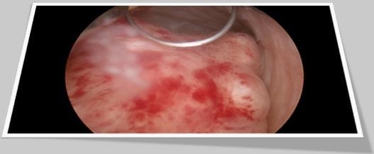
Hysteroscopy is a diagnostic and surgical procedure that makes examining the inside of the uterus possible without making an abdominal cut (incision). During hyteroscopy, a lighted viewing instrument called a hysteroscope is inserted through the vagina and cervix and into the uterus. Treatment can also be done through the hysteroscope during the same procedure.
Hysteroscopy usually takes 30 to 45 minutes. It is done as an outpatient procedure. General anesthesia is usually used, although local or spinal anesthesia can be used instead. You should not eat or drink for at least 4 to 8 hours before having the test. A gynecologist or surgeon does the procedure. The uterus is filled with a fluid, such as normal saline or glycine. The hysteroscope is inserted through the cervix into the uterus so the inner surface of the uterus can be examined. Hysteroscopy can locate the cause of bleeding for many women.
Hysteroscopy is used both to diagnose and treat abnormal vaginal bleeding. If areas of bleeding are found during the procedure, the tissue may be destroyed by laser beam or electric current (electrocautery) or surgically removed at the same time.
Hysteroscopy is done to Locate and evaluate the cause of uterine bleeding, such as uterine fibroids, when blood loss is severe.
Hysteroscopic management in submucous fibroids:
In the case of uterine submucous fibroid, infertility and obstetric complications are believed to be common compared with those with a normal uterine cavity. Over the last few years, hysteroscopy has allowed the transvaginal removal of submucous fibroids that are either prolapsed or located within the uterine cavity. Given the numerous advantages of the hysteroscopic approach over the transabdominal one, hysteroscopic myomectomy has become very popular. Our results are encouraging in terms of the reproductive outcome. Twenty-one women (72.4%) became pregnant after myomectomy. Thirteen women gave birth to 16 live infants. Nine of them delivered 12 viable term neonates. It is reported that 11 out of 31 infertile women (35.5%) became pregnant following hysteroscopic myomectomy with 9 term deliveries. A difference in delivery was found between patients with one submucous myoma resected than those with 2 or more. In the same way, it is concluded that hysteroscopic myomectomy appears to enhance fertility compared with infertile women with normal uterine cavity. The more recent study concerning that 25 women (60.9%) became pregnant with 48.7% live birth rate. Therefore, it suggests that an improved uterine contour may result in an improved pregnancy outcome and term deliveries in women with prior spontaneous pregnancy losses or primary infertility.
The possible role of submucous myomas in the causation of infertility is as yet unclear. It may be that in this case the cause of infertility is multifactorial. Meta-analysis has suggested also that removal of such tumors may be indicated in infertile women where no other factor has been identified. The relatively lower incidence of abortions and the fact that the rate of deliveries of live births in their first pregnancies increased significantly following treatment of the myomas, emphasize the major role of such fibroids as a cause of recurrent abortions and/or pregnancy losses and demonstrate the efficacy of the treatment.
Postoperative reproductive outcome is adversely affected by the presence of additional infertility factors. It is reported in their retrospective series that the pregnancy rate was 41.6% when the myoma was the only apparent cause, compared with 26.3% with the presence of one factor and 6.3% with two or more additional factors. In the current prospective study, none of our selected patients had additional infertility factors and this could explain the higher pregnancy rate reported in our series. These observations probably reinforce the fact that myomas could be a cause of infertility.
Conclusion:
In conclusion, it shows that hysteroscopic myomectomy clearly improves the rate of live birth for selected women with submucous myomas and history of primary infertility, recurrent abortion and/or preterm delivery. Ideally, in order to evaluate the efficency of this technique, randomized studies should be undertaken in multiple centers, taking into account a larger number of women suffering from this type of abnormality.


