How to Chose an Ideal High Definition Endoscopic Camera System
Telescope

Laparoscopy (from Greek lapara, "flank or loin", and skopein, "to see, view or examine") is an operation performed within the abdomen or pelvis through small incisions (usually 0.5-1.5 cm) with the aid of a camera. It may either be used to inspect and diagnose an ailment or to perform surgery. It can be used in endometriosis surgery. The telescope generally captures images via a distal-mounted objective lens. The image is then told through a rod-lens system to a proximal-mounted ocular lens that magnifies it for the surgeon. The resolution associated with a optical system is restricted to diffraction-point objects are converted into spots, and also the size of those spots is dependent upon the optical aperture, image magnification and lightweight wavelength. Generally, larger telescopes with lower magnification generate smaller spots, meaning higher resolution. Nevertheless, lens design and production quality are critical factors in determining resolution since suboptimal quality can help to eliminate image quality.
In laparoscope the lens diameter is very central determining resolution. For example, an expert HD camcorder having a 70mm lens along with a Ten times larger optical aperture provides significantly higher resolution images in contrast to the 10mm lens of an endoscope. The bigger lens is capable of transmitting a significantly larger bandwidth of information. Larger telescopes with lower magnification generate small spots, leading to higher resolution. Such optical characteristics imply that the perceived visual benefits of HD versus standard definition (SD) endoscopy are very subtle for many surgical specialties, though even smaller endoscopes displaying a full-circle image on a monitor may show a noticeably better image by having an HD camera.
Light Source

Now an eco-friendly LED Source of light reduces hazardous environmental waste by preventing frequent changes of sunshine bulbs. Being an added benefit, the not compulsory Safelight technology helps to ensure that the light source adopts standby mode once the Safelight cable detaches from the scope. This feature will lessen the risk of injury towards the patient and OR staff.
This LED light source is a great solution when used with the HD 3-Chip Camera as the light output settings are completely controllable from you head. Overall lower operating cost and increased safety get this to device a sure winner in the OR.
Light may be the essence of endoscopic imaging, and it is the starting point of the imaging chain. In the broadcast field, the quality of HD cameras is dependent on the performance characteristics of the lighting system being used, which is much the same with medical endoscopy. Endoscopy takes a high-performance source of light with plenty of intensity to make sure adequate illumination-even for light-critical applications. The illumination must ensure especially true-color properties and brilliant image presentation. The light sources must combine sufficient brightness and contrast to enable surgeons to tell apart healthy tissue from suspect tissue requiring treatment. Moreover, during therapeutic procedures, the sunshine source must adequately illuminate the operative field to allow the surgeon to obviously visualize anatomical structures and control the fragile movements of surgical instruments.
HD endoscopy generally places increased demands on surgical light sources. To get crisp, clear HD endoscopic images with superior resolution, optimum illumination must be provided to the operative field. Because HD cameras have lower sensitivity due to smaller pixel size, a powerful 300W Xenon light source is frequently recommended. Additionally, a chance to control surgical lighting, for example switching from active to standby modes, offers additional advantages to surgeons using HD imaging systems.
Camera Head
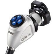
The camera head consists of a goal lens, a prism assembly and three sensors for acquiring the primary colors of the image. Some camera heads likewise incorporate an optical zoom for adjusting the look size (magnification). Optical zoom is advantageous because it doesn't have negative effect on the look resolution. In addition, a few of the image processing might be performed in the actual camera head. Due to better image performance, three-chip cameras were generally accepted as the industry standard for endoscopic surgery about Ten years ago. The primary advantage is the fact that color reproduction is much more natural.
In laparoscopic camera as with standard definition endoscopy systems, most HD Camera Heads acquire image data from the telescope. Image quality, however, will depend on the camera acquisition standard that's been put on a given HD system. The main difference between SD and HD video formats is better visualized by comparing the vertical and horizontal resolution. Typical SD formats offer a 4:3 aspect ratio, 640 by 480 horizontal and vertical lines. The HD format provides a 16:9 aspect ratio, 1280 by 720 horizontal and vertical lines. The 1080 HD standard also offers a 16:9 aspect ratio, but 1920 by 1080 horizontal and vertical lines-seven times the SD resolution at 480 lines. The temporal resolution may be the quantity of captured images expressed as frames per second (fps). The 720p standard represents progressive scanning-capturing the whole frame as you image 60 times per second. The 1080i standard represents interlaced scanning-capturing two fields of half images with alternating lines that are then combined to produce each complete frame.
In laparoscopic operations for HD endoscopy, 1080p60 (1080p at 60fps) may be the highest standard readily available for acquiring and displaying images, while offering a superior viewing experience for surgeons. Because progressive scanning offers twice the temporal resolution when compared with interlaced scanning, it is well suited for fast paced objects and capturing still images, developing a quasi-3D image. Anatomical structures become visible which were hidden in the flatter SD image.
Almost like other optical systems in entertainment tachnology, video camera lenses create circular images. Nevertheless, movies, shows and sports events are viewed on the broadcast HDTV set with a wide-screen 16:9 aspect ratio. This is accomplished by over framing the look so that the wide-screen sensor is totally taught in circular optical image (see image on “Peripheral Vision”). The same is true for laparoscopic surgery where surgeons watch a monitor screen completely filled through the surgical image. The adjacent image (“peripheral vision”) demonstrates the problem for 2 different aspect ratios. Wide-screen image acquisition increases the horizontal field of view (panoramic image) and decreases the vertical field of view. With laparoscopic instruments primarily entering the concept of view laterally, a wide-screen 16:9 aspect ratio seems advantageous. Pulling back the telescope slightly can make amends for losing in vertical field of view. Another positive effect is the fact that a telescope positioned further in the site of surgical interaction catches less debris and smoke on the front window, improving image quality.
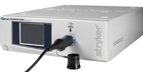
Camera CCU
An Ideal HD 3-Chip Endoscopic Camera is a groundbreaking new development due to its ability to allow highest resolution within the operating room when associated with hd HD monitor. With the HD technology. HD 3-Chip Camera should includes a 1920x1080p resolution giving the surgeon the command to control the sunshine source settings from your camera head. Also, by getting for backwards compatibility with past generation digital capture devices, scopes and various flat panels, the 1288 HD 3-Chip Camera is constantly on the offer new solutions for improved visualization within the operating room.
The Camera CCU connects various aspects of the HD imaging chain, capturing and processing video signals from the camera head for display about the monitor, as well as for transfer to existing recording and printing devices. In addition to processing digital HD images, the Camera CCU should be able to either down-converting HD signals to SD or up-converting SD signals to HD. This allows SD printers and capture units to continue getting used with the HD system. Additionally, it allows images in one format to become viewed, with some limitations, on displays of the different format. In this way, compatibility of the HD system with both SD and HD components extends the capabilities of facilities that still rely on SD components for some applications. In typical surgical settings, a mixture of SD and HD imaging components is likely to be found. The HD Camera CCU must offer flexible output choices to ensure that the unit can continue to be used with HD equipment. The CCU should be able to accommodate both SD and HD input and, conversely, it will have two digital video outputs-DVI (digital video interface) for the HD signal, and SDI (serial digital interface) for the SD signal.
Video Cables
Video cable has a great importance in video system of minimal access surgery. Video Cables that carry digital image data between the camera head, camera CCU, and monitor and recording devices must offer sufficient bandwidth for video transmission of HD data over long distances. For example, a 1080p HD imaging system provides approximately 2 million pixel resolution at 60 frames per second, requiring twice the bandwidth like a 1080i system at 30 fps. Using the introduction of optical fiber, DVI cabling can now achieve this purpose. It represents the simplest way of transmitting a 1080p signal. Essentially, the introduction of optical fiber provides an optimum cable solution for transmitting HD signals over long distances. This gives a chance to transmit other HD signals from imaging sources such as PACS.
An ideal optical fiber for HD signal transmission can also be necessary for the development of HD imaging technology into integrated operating room systems. Depending on the level of system integration anticipated for any new OR, a distribution amplifier or matrix router may be needed to provide necessary DVI transmission and distribution functionality. Where HD capabilities are now being put into existing integrated operating rooms, chances are that rewiring will be necessary to ensure HD compatibility of all cabling in the room.
Monitor

A 26” HD monitor provides clear, brilliant images on a spacious, widescreen display. Offering both 4:3 and 16:9 aspect ratio display formats, improved color reproduction and new picture-by-picture capabilities. An ideal high definition platform is designed to create the optimal operating environment for the surgeon and gynecologist. A Wide-Screen HD Monitor displaying images acquired in 16:9 format enables surgeons to experience more natural vision. More importantly, visualization is much more in tune with human anatomy.
In minimal access surgery to achieve the full benefit of HD imaging and maximize performance, the monitor resolution must be properly matched to the camera head acquisition resolution. High-quality HD flat-panel monitors display image data like a progressive scan. Which means that a 1080i HD signal should be deinterlaced to complement the monitor format, along with a 720p HD signal might be up-converted to have an HD monitor with 1200 vertical lines. On the other hand, a 1080p HD signal requires no conversion for any 1920x1200 HD monitor if small black bars at bottom and top are acceptable.
In laparoscopic surgery a significant benefit of 16:9 HD monitors would be that the images offer a natural, panoramic view. In humans, our horizontal field of view is wider than our vertical field of view. For surgeons, this wider, natural view is less fatiguing during procedures. Additionally, during laparoscopic surgery as surgeons are viewing full-screen endoscopic images, trocars and hand instruments that normally approach the surgical area laterally is visible earlier with a 16:9 monitor than they are able to on the 4:3 or 5:4 monitor.


