"Fibroids" or "uterine myomas (short for leiomyoma)", 30% of women are influenced by it. The terms "fibroid" and "myoma" are synonyms. Most fibroids do not cause symptoms, and do not require treatment.
Treatment may require for Fibroids in the following conditions:
- Fibroids are growing large can cause pressure on other organs, bladder is an example.
- Rapid growth of Fibroids
- Abnormal bleeding caused by Fibroids
- Fertility related problems caused by Fibroids
Kinds of Fibroids:
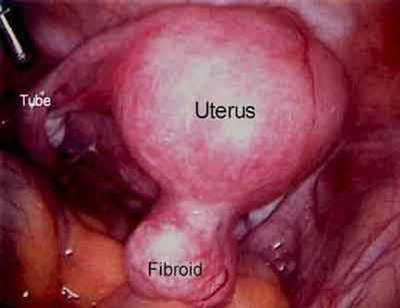
Basis of classification of Fibroids done by their location (see figure above), which influences the symptoms they may cause and may influence mode of treatment. Fibroids that are inside the cavity of the uterus will usually cause bleeding between periods (metrorrhagia) and often cause severe cramping. Fortunately, a method called "hysteroscopic resection,” by which fibroids can usually and easily be removed, which can be done through the cervix without the need for an incision. Presence of Sub mucous myomas are detected partially in the cavity and partially in the wall of the uterus. Heavy menstrual periods can be caused (menorrhagia), bleeding between periods as well. Some of these can also be removed by hysteroscopic resection.
Intramural myomas are found in the wall of the uterus, and ranging in size from microscopic to larger than a grapefruit. Unless they become quite large, many of these do not cause problems. There are a number of alternatives for treating these, but often they do not need any treatment at all. The outside wall of the uterus, Subserous myomas are commonly found, and may even be connected to the uterus by a tail (pedunculated myoma.) unless they grow large, these do not need treatment, but those on a tail can twist and cause pain. To remove by laparoscopy, this kind of fibroid is the easiest.
Diagnosis of Fibroids
During a pelvic exam Fibroids may be felt, if the examiner relies just on the examination, many times myomas that are causing symptoms may be missed. Also, other conditions such as adenomyosis or ovarian cysts may be mistaken for fibroids. For this reason, when a woman has symptoms of abnormal bleeding or cramping, routinely doing an ultrasound examination at the time of the first visit is essential, or if any abnormality felt on examination. Only takes a few minutes to do Vaginal probe ultrasound, is not uncomfortable, and rapidly provides helpful information if the examiner is experienced in looking at uterine abnormalities. During the ultrasound, it is possible to fill the uterus with a liquid (saline enhanced sonography or sonohysterogrami). While this will often provide additional information to the regular ultrasound, with a little telescope one can usually learn much more by looking inside the uterus. This exam, called hysteroscopy, is usually a quick office procedure that allows directly looking inside the uterus.
Adenomyosis is One of the most common conditions confused with fibroids. The lining of the uterus infiltrates the wall of the uterus in adenomyosis, causing the wall to thicken and the uterus to enlarge. On ultrasound examination, this will often appear as diffuse thickening of the wall, while fibroids are seen as round areas with a discrete border. Adenomyosis is usually a diffuse process, and without taking out the uterus, rarely can be removed. Since fibroids can be removed or treated by uterine artery embolization, before planning treatment, it is important to distinguish between the two conditions. In addition to fibroids it is also common to have some adenomyosis.
An excellent picture of the uterus made available by MRI scans. An MRI provides thorough pictures of the uterus and fibroids, and can off tell the dissimilarity between adenomyosis and fibroids.


