Equipment detail 1, Equipment detail 2, Equipment detail 3, Equipment detail 4, Equipment detail 5
READ ABOUT LAPAROSCOPIC EQUIPMENT AND INSTRUMENTS
Laproflattor
The Electronic CO2 Laproflattor is a general purpose insufflation unit for use in laparoscopic examinations and operations. Controlled pressure insufflation of the peritoneal cavity is used to achieve the necessary work space for laparoscopic surgery by distending the antero-lateral abdominal wall and depressing the hollow organs and soft tissues. Carbon dioxide is the preferred gas because it does not support combustion, it is very soluble which reduces the risk of gas embolism, and is cheap. Automatic insufflators allow the surgeon to preset the insufflating pressure, and the device supplies gas until the required intra-abdominal pressure is reached. The insufflator activates and delivers gas automatically when the intra-abdominal pressure falls because of gas escape or leakage from the ports. The required values for pressure and flow can be set exactly using jog keys and digital displays. Insufflation pressure can be continuously varied from 0 to 30 mm Hg; total gas flow volumes can be set to any value in the range 0-9.9 liters/mm.

Figure 2-28: Insufflator
Patient safety is ensured by optical and acoustic alarms as well as several mutually independent safety circuits. The detail function and quadro-manometric indicators of insufflator is important to understand safety point of view. The important indicators of insufflators are preset pressure, actual pressure, flow rate and total gas used.
Suction Irrigation Machine or Pelvis-Cleaner
Figure 2-29: Laparoscopic Suction Irrigation Machine
It is used for flushing the abdominal cavity and cleaning during endoscopic operative intrusions. It has been designed for use with the 26173 AR suction /instillation tube. Its electrically driven pressure/suction pump is protected against entry of bodily secretions. The suction irrigation machine is used frequently at the time of laparoscopy to make the field of vision clear. Most of the surgeons use normal saline or ringer lactate for irrigation purposes. Sometime heparinized saline is used to dissolve blood clot to facilitate proper suction in case of excessive intra-abdominal bleeding.
Disposable or Reusable Instrument
Several factors should be considered at the time of choosing laparoscopic instrument, including cost, availability and reliability. Reusable instruments are expensive initially but in long rum they are cost effective. The disposable instrument cost is less compared to re-usable but patient cost is increased. In many centers re-use of disposable instrument is seen. In developing countries, disposable instruments are very rarely used because labour cost is low compare to the cost of disposable instrument. In Europe and USA, surgeons often choose to use disposable instrument in order to save high labour cost. The main advantage of disposable instrument is high performance due to its sharpness and reduced chance of disease transmission due to certified high-end factory sterilization. However, once discarded, environment concerns are raised about disposal and biodegrability of disposable instruments. Ideally disposable instrument should not be used repeatedly because handling, sorting, storing and sterilization make these instrument questionable. The disposable instruments are not sterilized properly by dipping in gluteraldehyde because they are not dismountable. Insulation of disposable instrument also can be torn easily which can lead to electrosurgical injuries.
Veress Needle
Veress needle was invented by a chest physician for aspiration of pleural effusion keeping in mind that its spring mechanism and blunt tip will prevent the injury of lung tissue. Veress needle consists of an outer cannula with a beveled needle point for cutting through tissues. Inside the cannula is an inner stylet, which is loaded with a spring that spring forward in response to the sudden decrease in pressure encountered upon crossing the abdominal wall and entering the peritoneal cavity. The lateral hole on this stylet enables CO2 gas to be delivered intra-abdominally.
Veress needle is used for creating initial pneumoperitoneum so that the trocar can enter safely and the distance of abdominal wall from the abdominal viscera should increase. Veress needle technique is the most widely practiced way of access. Before using veress needle every time it should be checked for its potency and spring action. Veress needle is available in three length 80mm, 100mm, 120mm. In obese patient 120mm and in very thin patient with scaphoid abdomen 80mm veress needle should be used. Veress needle should be held like a dart at the time of insertion. The proper technique of veress needle insertion, different safety measures and indicators are discussed later in access technique.
Trocar and Cannula
The word "trocar" is usually used to refer to the entire assembly but actual trocar is a stylet which is introduced through the cannula. The trocars are available with different type of tips. The cutting tips of these trocars are either in the shape of a three edged pyramid or a flat two edged blade
Conical tipped trocars are supposed to be less traumatic to the tissue. The tip can be penetrated through the parietal wall without cutting and decreased risk of herniation or haemorrhage is reported.
Figure 2-32: Disposable Trocar and Cannula
Cannulas are in general made from plastic or metal. Plastic devices whether they are transparent or opaque, need to be designed in such a way as to minimize the reflection of light from the telescope. Reusable and disposable trocars are constructed by a combination of metal and plastic. The tip of disposable trocar has a two edged blade. These are very effective at penetrating the abdominal wall by cutting the tissue as they pass through. Most of the disposable plastic trocar has a spring loaded mechanism that withdraws the sharp tip immediately after it passes through the abdominal wall to reduce the incidence of injury of viscera. Trocar and cannula are of different sizes and diameter depending upon the instrument for which it is used. The diameter of cannula ranges from 3 mm to 30 mm; the most common size is 5mm and 10 mm. The metal trocar has different type of tips i.e. pyramidal tip, Eccentric tip, conical tip or blunt tip depending on the surgeon's experience.
All the cannula has valve mechanism at the top. Valves of cannula provide internal air seals, which allow instruments to move in and out within cannula without the loss of pneumoperitoneum. These valves can be oblique, transverse, or in piston configuration.
These valves can be manually or automatically retractable during instrument passage. Trumpet type valves are also present which provide excellent seals, but they are not as practical as some of the other systems. They require both hands during instrument insertion, which may explain why they are less often used in advanced laparoscopic cases. The flexible valves limit the carbon dioxide leaks during work whatever the diameter of the instrument used.
Surgeon should remember that sharp trocars although looking dangerous are actually better than blunt one because they need less force to introduce inside the abdominal cavity and chances of inadvertent forceful entry of full length of trocar is less. There is always a difference in the marked exterior diameter of the cannula and the interior usable diameter. The end of the cannula is either straight or oblique. An oblique tip is felt to facilitate the easy passage of the trocar through the abdominal wall.
Trocar and cannula should be held in proper way in hand so that head of the trocar should rest on the thenareminence, the middle finger should rest over the gas inlet and index finger is pointed towards the sharp end of the trocar.
Laparoscopic Hand Instruments
Laparoscopic hand instruments vary in diameter from 1.8 to 12mm but majority of instruments are designed to pass through 5 to 10mm of cannula. The hand instrument used in laparoscopic surgery are of different length (varies company to company and length of laparoscopic instrument varies from 18 to 45cm) but they are ergonomically convenient to work if they have same length of approximately 36 cm in adult and 28 cm in pediatric practice. Shorter instruments 18 to 25cm are adapted for cervical and pediatric surgery. Certain procedures for adult can also be performed with shorter instrument where the space is constricted. 45cm instruments are used in obese or very tall patients. For better ergonomics half of the instruments should be inside the abdomen and half outside. If half of the instrument is in and half out, it behaves like class 1 lever and it stabilizes the port nicely so the surgery will be convenient.
Most of the laparoscopic procedures require a mixture of sharp and blunt dissection techniques, often using the same instrument in a number of different ways. Many laparoscopic instruments are available in both re-usable and disposable version. Most re-usable instruments are partially dismountable so that it can be cleaned and washes properly. Some manufacturer have produced modular system where part of the instrument can be changed to suit the surgeon favorite attachment like handle or working tip.
Most laparoscopic instruments like graspers and scissors has basic opening and closing function. Many instrument manufacturers during past few years are able to rotate at 360 degree angle which increases the degree of freedom of these instruments.
Certain types of instrument offer angulations at their tip in addition to usual 4 degree of freedom. These instruments are used to avoid obstacles and for the lateral grasping when the instrument is placed outside of the visual field. This feature is available for both re-usable as well as disposable instrument. The complex mechanism of such instrument makes their sterilization very difficult.
A variety of instruments, especially retractors have been developed with multiple articulations along the shaft. When these are fixed with the tightened cable the instrument assumes a rigid shape which could not have been introduced through the cannula.
Most of the hand instrument has three detachable parts.
- Handle
- Insulated outer tube and
- Insert which makes the tip of the instrument
Different Handles of Hand instrument
Certain instruments handle are designed to allow locking of the jaw. This can be very useful when the tissue needs to be grasped firmly for long period of time preventing the surgeons hand from getting fatigue. The locking mechanism is usually incorporated into the handle so that surgeon can easily lock or release the jaws. These systems usually have a ratchet so that the jaws can be closed in different position and to different pressure. Most of the Laparoscopic instruments handle has attachments for unipolar electrosurgical lead and many have rotator mechanism to rotate the tip of the instrument. Some multifunctional Laparoscopic handle has attachment for suction and irrigation and some time hand switch for cutting and coagulation switch of electrosurgery.
Cuschieri Ball Handle is invented by Prof. Sir Alfred Cuschieri. This handle lies comfortably in surgeon's palm. This design reduces the fatigue of surgeon and eases rotation of the instrument by allowing rotation within the palm rather than using wrist rotation. Squeezing the front of the handle between the thumb and the first fingers increases the jaw closing force; squeezing the rear of the handle between the eminence of the thumb and last fingers opens the jaws.
Cuschieri pencil handle also has great ergonomic value specially when used with needle holder. This handle allows the angle between the handle and the instrument to be altered to suit the surgeon's wrist angle. The conveniently placed lever of this pencil handle when pressed can change the angle. Just like ball handle, pressure at the front increases the jaw closing force while pressure at the rear opens the jaw.
Outer Sheath of Hand Instrument
The insulation covering of outer sheath of hand instrument should be of good quality in hand instrument to prevent accidental electric burn to bowel or other viscera.
Insulation covering may be of silicon or plastic. At the time of cleaning the hand instrument, utmost care should be taken so that insulation should not be scratched with any sharp contact. A pin hole breach in insulation is not easily seen by naked eye but may be dangerous at the time of electro surgery.
Insert of Hand Instrument
Insert of hand instrument varies only at tip. It may be grasper, scissors, or forceps. This grasper may have single action jaw or double action jaw. Single action jaw open less than double action jaw but close with greater force thus, most of the needle holders are single action jaw. The necessary wider opening in double action jaw is present in grasper and dissecting forceps. Single action graspers and dissectors are used where more force is required.
Single Action Jaw Graspers
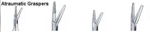
These graspers are good when you don't have control over depth and surgeon wants to work in single plane in controlled manner particularly during adhesiolysis.
Double Action Jaw Graspers

Instruments for Sharp Dissection
- Scissors
- Electro surgery hook
- HF Electro surgery spatula (Berci)
- HF Electro surgery knife
- Knife
Scissors

Figure 2-49: Disposable
ScissorsJean-Claude Margueron of Emar in Fourteenth Century B.C. invented scissors. Scissors are one of the oldest surgical instruments used by surgeons. Scissors are used to perform many tasks in open surgical procedure but its use in minimal access surgery is restricted. In minimal access surgery scissors require greater skill because in inexperienced hand it may cause unnecessary bleeding and damage to important structures.
Mechanism of cutting:
The scissors has three parts:
- Blade
- Fulcrum
- Handle
The cutting force of the scissors works on the law of lever. The force applied on the blade can be calculated by length of the handle and force applied on the grip of handle. A pair of scissors is an example of first class levers connected together at the joint known as fulcrum.
There are three type of lever:
Scissors works on the principle of class 1 lever. In class 1 Lever, the pivot (fulcrum) is between the effort and the load. The more the length of the handle or the fulcrum of the scissors, the less force of cutting will be required. The laparoscopic scissors do not apply the exact law of lever because of the cylinder action of the long shaft, but the design of handle helps in the amplification of force by lever action.
Scissors function by the combination of:
- Gripping
- Squeezing
- Tearing
When the blades of scissors close, its sharp edges grind against each other and any tissue which comes between the blades of scissors will get cut. The scissors-tissue interaction can be described in five stages:
1.Engagement
In the process of engagement, the two blades of the scissors engage a piece of tissue to cut. The amount of tissue engaged should not be more than the space between the jaw of blades otherwise the chance of slipping of tissue is more. After engagement, the force applied on the handle of the scissors initiate cutting.
2. Elastic deformation
This stage starts just after the engagement of tissue between the blades of the scissors. In this process, the tissues between the two blades of scissor start deforming. This stage is called elastic deformation, because if the force on the handle of scissors is removed then the tissue deformity will return to its normal state.
3. Plastic deformation
Further force on the handle of scissors will cause the tissue between the blades to go into a plastic deformed state, which is irreversible. After undergoing this state of tissue deformation, even if further process of cutting is stopped the impression on the tissue remains.
4. Fracture
Further increased force on the fulcrum of scissors will result in the fracture of intercellular plane of the tissue. This stage of cutting is peculiar to scissors because unlike the scalpel, the site of tissue fracture is intercellular.
5. Separation
After the fracture the tissue separates along line of the blade of scissors, and then this whole process of cutting will continue on the engaged tissue.
Histology of the tissue after cutting
Histological examination of the tissue after cutting with scissors shows that there is separation of tissue through intra cellular plane. Microscopic examination shows serrated cut margin along the line of tissue separation.
Types of Laparoscopic Scissors
Straight Scissors
The blade of this scissor is straight and it is widely used as an instrument for mechanical dissection in laparoscopic surgery. Straight scissor can give controlled depth of cutting because it has only one moving jaw. At the time of cutting the fixed jaw should be down and moving jaw should be up.
Curved Scissors
The blade of this scissors is slightly curved and this is the most widely used scissor in laparoscopic surgery. These scissors are mounted on a curved handle which is either fixed or retractable. The type with a fixed curvature proximal to the scissor blades require introduction through flexible valve-less ports. The surgeon prefers this scissors because the curvature of the blade of this scissors abolishes the angle of laparoscopic instruments manipulation and better view through telescope.
Serrated Scissors
The main advantage of this scissors is that the serrated edges prevent the tissue to slip out of the blades. It is a useful instrument in cutting a slippery tissue or ligature. Serrated scissors may be straight or curve.
Hook Scissors
The sharp edge of both blades is in the shape of a flattened C. The blades can be partially closed, trapping tissue in the hollow of the blades without dividing it and allowing it to be slightly retracted. This allows the surgeon to double check before he closes the blades completely.
The main advantage of this scissors is that, it encircles the structure before cutting: Tissue is held between its jaws and there is no chance of slipping. The Hook scissor is especially useful for cutting secured duct or artery in laparoscopic surgery. The cutting of nerve bundle in neurectomy becomes very easy with the help of this scissors. Hook scissors is also helpful in partial cutting of cystic duct for intra-operative Cholangiography. All the other scissors cut from proximal to distal whereas the hook scissors cut distal to proximal
Micro-tip Scissors
These very fine scissors, are either straight or angled, and are used to partially transect the cystic duct. The main advantage of this scissors is to cut the ducts partially for facilitating cannulation. This scissor may be used for cutting the cystic duct for performing intra-operative Cholangiogram. Exploration of small ducts like common bile duct is very helpful with micro scissors due to its fine small blades. Fine micro scissors are also available in its curved form.
The use of scissors endoscopically requires little modification of open techniques. The basic instrument is a miniaturized, long handled version of conventional scissors and can be single or double action. There are some special types of scissors used in endoscopic surgery.
Test your knowledge with laparoscopic quiz
Subscribe your free monthly journal of Laparoscopy

Download prospectus of Laparoscopic Training
READ ABOUT LAPAROSCOPIC EQUIPMENT AND INSTRUMENTS
Few among hundred of Surgeons & Gynaecologists trained at our Hospital


