Laparoscopic Myomectomy: Stepwise Approach to Large Posterior Wall Intramural Fibroids
Add to
Share
362 views
Report
4 months ago
Description
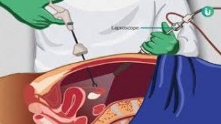
Laparoscopic myomectomy has emerged as a gold standard for the management of uterine fibroids, offering patients the advantages of minimal invasiveness, reduced postoperative pain, faster recovery, and superior cosmetic outcomes compared to traditional open surgery. The surgical removal of large posterior wall intramural fibroids, however, poses unique technical challenges due to their size, location, and proximity to vital uterine structures. A meticulous step-by-step approach is essential to ensure surgical safety, minimize complications, and preserve uterine integrity for future fertility. Preoperative Preparation Successful laparoscopic myomectomy begins with thorough preoperative planning. Detailed imaging, typically via transvaginal or pelvic ultrasonography and magnetic resonance imaging (MRI), helps define the size, number, and location of fibroids. Patients are counseled regarding the benefits and risks of the procedure, including possible conversion to open surgery if intraoperative difficulties arise. Preoperative medical therapy, such as gonadotropin-releasing hormone (GnRH) analogs, may be considered to reduce fibroid size and vascularity, although this is tailored to individual cases. Step 1: Patient Positioning and Port Placement The patient is placed in the lithotomy position with a slight Trendelenburg tilt to facilitate visualization of the pelvic organs. After induction of general anesthesia, pneumoperitoneum is created, typically through a Veress needle or open technique, and a primary camera port is inserted. Additional accessory ports are strategically placed under direct visualization to allow optimal instrument maneuverability, especially for accessing the posterior uterine wall. Step 2: Visualization and Uterine Manipulation A uterine manipulator is used to provide mobility and improve exposure of the posterior wall. The peritoneal cavity is carefully inspected, and the fibroid’s relationship with surrounding structures, including the bladder, bowel, and major vessels, is assessed. Step 3: Incision and Fibroid Enucleation A serosal incision is made over the most prominent part of the fibroid using monopolar scissors or harmonic energy, taking care to minimize bleeding. Vasoconstrictive agents, such as diluted vasopressin, can be injected into the myometrium to reduce intraoperative blood loss. Blunt and sharp dissection is employed to enucleate the fibroid, carefully maintaining the integrity of the surrounding myometrium. For large posterior wall intramural fibroids, meticulous traction-countertraction techniques are critical to facilitate complete removal. Step 4: Hemostasis and Myometrial Closure After enucleation, meticulous hemostasis is ensured. The myometrial defect is closed in multiple layers using barbed or absorbable sutures to restore uterine wall strength and prevent postoperative complications such as hematoma formation or uterine rupture in future pregnancies. Step 5: Specimen Retrieval The excised fibroid is morcellated, either manually or with contained power morcellation techniques, for safe extraction through laparoscopic ports. Surgeons must adhere to current safety guidelines to minimize tissue dissemination. Step 6: Peritoneal Irrigation and Closure The peritoneal cavity is thoroughly irrigated to remove any blood or debris, and the laparoscopic ports are removed under direct vision. Port sites are closed appropriately, ensuring minimal risk of herniation or infection. Postoperative Care Patients are monitored for hemodynamic stability and postoperative pain. Early ambulation and gradual resumption of diet are encouraged. Recovery is generally rapid, with most patients resuming normal activities within one to two weeks. Conclusion Laparoscopic myomectomy for large posterior wall intramural fibroids is a technically demanding but highly effective procedure. Mastery of this technique requires a combination of detailed preoperative planning, refined surgical skills, and adherence to a step-by-step approach. When performed meticulously, it offers patients the dual benefits of effective fibroid removal and preservation of uterine function, making it the preferred choice for women seeking fertility preservation and minimal surgical morbidity.
Similar Videos