Bleeding in Gallbladder Bed: Laparoscopic Hemostasis Techniques
Add to
Share
1,009 views
Report
7 months ago
Description
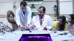
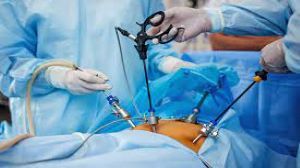
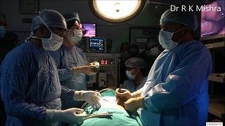
Bleeding from the gallbladder bed is one of the most common intraoperative challenges encountered during laparoscopic cholecystectomy. At World Laparoscopy Hospital (WLH), surgeons are trained to manage this complication with precision, patience, and advanced laparoscopic techniques to ensure complete hemostasis while maintaining tissue safety. Understanding the Source of Bleeding During laparoscopic cholecystectomy, bleeding may occur from: The cystic artery stump, if not adequately clipped or coagulated. The liver parenchyma at the gallbladder bed after dissection. Small venous channels running between the gallbladder and liver. Recognizing the source promptly is crucial for effective management and avoiding postoperative hemorrhage or bile leakage. Step-by-Step Approach to Control Bleeding at WLH At World Laparoscopy Hospital, surgeons follow a structured, evidence-based protocol to control bleeding safely: 1. Clear Visualization The first rule is not to panic. The laparoscope lens is cleaned, and suction is used to clear pooled blood for an unobstructed view. The surgeon ensures proper insufflation pressure (12–14 mmHg) to maintain a clear operative field. 2. Identify the Source The bleeding point is carefully identified using gentle irrigation and suction. Avoid aggressive suctioning that might enlarge a venous tear or dislodge a forming clot. 3. Use of Energy Devices WLH emphasizes energy-based hemostasis using advanced instruments like the Harmonic Scalpel, LigaSure, or bipolar coagulation. These devices seal small vessels efficiently with minimal thermal spread, preserving liver tissue integrity. 4. Pressure and Packing Technique For diffuse oozing from the liver bed, a gentle pressure tamponade using a piece of gauze or Surgicel is applied for 3–5 minutes. This simple yet effective technique allows small vessels to coagulate naturally. 5. Topical Hemostatic Agents WLH surgeons are trained to use oxidized regenerated cellulose, fibrin glue, or gelatin sponges over the gallbladder fossa when oozing persists despite cautery. These agents enhance clot formation and minimize the risk of delayed bleeding. 6. Clip or Suture if Required If a spurting artery is identified, a laparoscopic clip or transfixation suture is applied using a 5-mm needle holder. Precise suturing skills, taught extensively in WLH training programs, are vital for such scenarios. 7. Irrigation and Final Inspection After achieving hemostasis, the area is irrigated with warm saline to remove blood clots. The surgeon inspects the Calot’s triangle and gallbladder bed thoroughly before closing, ensuring a completely dry field. Postoperative Precautions A closed suction drain may be placed if there was significant bleeding during surgery. Patients are closely monitored for tachycardia, falling hemoglobin, or abdominal distension, which could indicate postoperative bleeding. Training Excellence at World Laparoscopy Hospital At WLH, under the mentorship of Dr. R. K. Mishra, surgeons learn these advanced hemostatic techniques through hands-on simulation, live surgeries, and structured skill labs. The emphasis is on safe dissection, gentle tissue handling, and proactive bleeding control—cornerstones of modern laparoscopic surgery. Conclusion Controlling bleeding in the gallbladder bed requires a calm, methodical approach, technical proficiency, and deep anatomical understanding. At World Laparoscopy Hospital, surgeons are not only trained to manage such complications effectively but also to prevent them through precise surgical planning and advanced laparoscopic skill. The result is a safer surgery, faster recovery, and a new standard of minimally invasive excellence.
Similar Videos